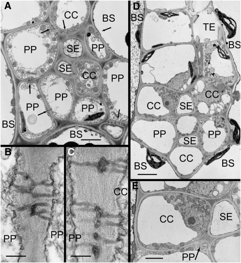
Figure 1.
Electron micrographs of apple (A and B) and P. major (C–E) minor veins in transverse sections. A, SEs and CCs are surrounded by PP cells. Numerous plasmodesmata are present at BS-PP and PP-CC interfaces (arrows), providing symplastic continuity into the phloem. B, Plasmodesmata between PP cells. C, Plasmodesmata between PP cells and CCs. D, SEs are surrounded by a ring of CCs and PP cells. Few plasmodesmata are visible at any interface. E, PP cells have transfer cell wall ingrowths (arrow) at the interfaces with SEs and CCs. Scale bars: A and E = 2 μm; B and C = 0.2 μm; D = 4 μm.

