
FIGURE 9. Western immunoblot analysis of cellular fractions of S. aureus Newman and Newman spa for IgG binding.
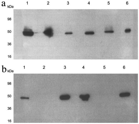
Cell envelopes of S. aureus Newman (a) or Newman spa (b) were separated into cell wall (lane 2), protoplasts (lane 3), protoplast membranes (lane 4), and cytoplasm fractions (lane 5) and analyzed by SDS-PAGE. A whole cell lysate (lane 1) and a concentrated supernatant representing secreted proteins (lane 6) were also included. Gels were analyzed by Western blotting with peroxidase-conjugated rabbit IgG. Approximate molecular weights are indicated.