
Figure 1.
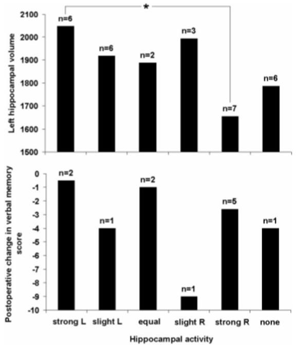
Categorization of hippocampal activity by visual inspection: The upper panel shows the relationship between visually determined hippocampal activity and left hippocampal volume (mm3, corrected for total intracranial volume); the lower panel shows the relationship between these categories and postoperative change in delayed recall of previously learned word lists. The visually determined categories are as follows: strong L (R) = strongly left (right) predominant activity; slight L (R) = slightly predominant activity on the left (right); equal activity on the two sides; no activity evident at p = 0.05. The asterisk shows a significant difference in left hippocampal volume for the strongly left and right lateral ized groups at p = 0.05. n = number of patients in each category.