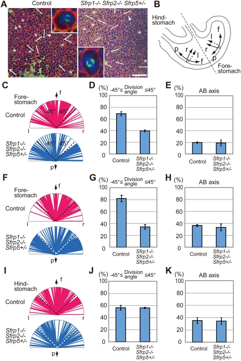
Figure 2. Divergence of cell division orientation in the epithelium of Sfrps-deficient fore-stomach.
(A) Cell division orientation was visualized with anti-ß1-integrin (red), anti-acetylated α-tubulin (green) and DAPI (blue) staining in the greater curvature epithelium of the fore-stomach of control and Sfrp1−/− Sfrp2−/− Sfrp5+/− embryos at E13.5. The top of the confocal image is oriented in the direction of the fundus. Scale bar: 40 µm. An inset shows higher magnification of the mitotic cells indicated by an arrowhead. The cell division axis is indicated by a bar, whereas the vertical axis is denoted by an asterisk. (B) Arrows indicate the direction of the fundus (f), pylorus (p) as well as right (r) and left (l) in the schematic diagram of the stomach at E12.5∼E13.5. (C–H) Cell division orientation converged within ±45° of the cephalocaudal axis in control fore-stomach epithelium, whereas it diverged in Sfrp1−/− Sfrp2−/− Sfrp5+/− fore-stomach epithelium at E12.5 (C, D) and E13.5 (F, G). Statistical analysis revealed a significant difference in convergence of cell division orientation along the cephalocaudal axis between control and Sfrps-deficient fore-stomachs at E12.5 (D) and E13.5 (G). In contrast, no significant difference in the frequency of cell division along the AB axis is evident between control and Sfrp1−/− Sfrp2−/− Sfrp5+/− fore-stomachs at E12.5 (E) and E13.5 (H). (I, J) Oriented cell division is not observed in hind-stomach epithelium of controls or Sfrp1−/− Sfrp2−/− Sfrp5+/− mutants at E13.5. (K) Cell division along the AB axis occurs at a similar frequency in control (35.1±4.93%, n = 3) and Sfrp1−/− Sfrp2−/− Sfrp5+/− (34.3±5.5%, n = 3) hind-stomachs at E13.5.

