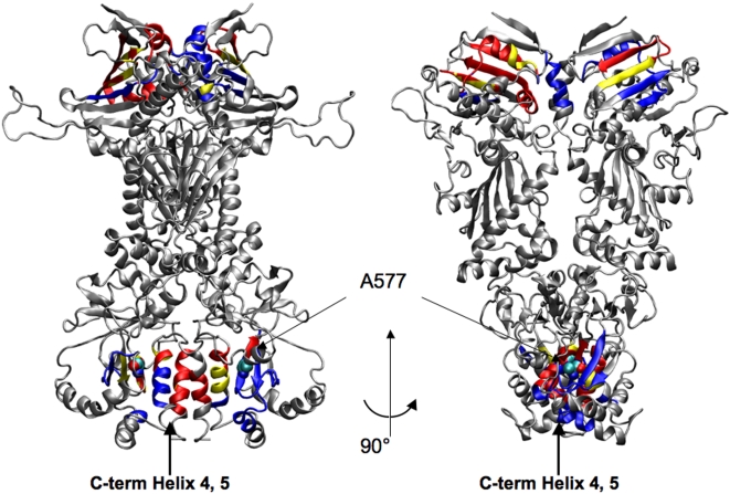
Figure 14. Representation on the 3D structure of residues communicating with high efficiency.
Two views of Hsp90 dimer, where residues active in very long range signalling (i.e. active in communication with other residues that are more than 80 Angstroms apart) are coloured according to the following scheme: Yellow: Residues active both in ATP and ADP system. Red: Residues active in the ATP system. Blue: Residues active in the ADP system. The mutation site A577 leading to loss of the ATPase activity is highlighted with spheres and labelled.

