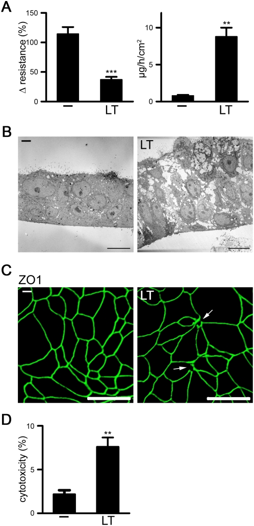
Figure 1. LT destroys barrier function in differentiated mucociliary human lung epithelium.
(A–D) Polarized NHBE were treated with or without LT for 48 h. (A) The epithelial layer was subjected to resistance measurements (left panel) or incubated with FITC-albumin on the apical side for permeability measurements (right panel). Data are represented as mean+/−SEM of 3–5 inserts/per condition, n = 3. (B) TEM images of crosscuts obtained from untreated (-) and LT-treated lung epithelial layers. Crosscuts with ciliated cells on the apical side are depicted. Scale bar represents 10 µm. (C) Confocal images of the apical side of untreated and LT-treated lung epithelial layers using a tight junction marker (ZO1, green). Z-series was analyzed with Imaris5 (Bitplane Inc., MN). Disruptions of multicellular junctions are indicated by white arrows. The scale bar represents 50 µm. (D) LDH release of untreated and LT-treated NHBE 3D layers. Data are represented as mean+/−SEM of 3 inserts, n = 3.

