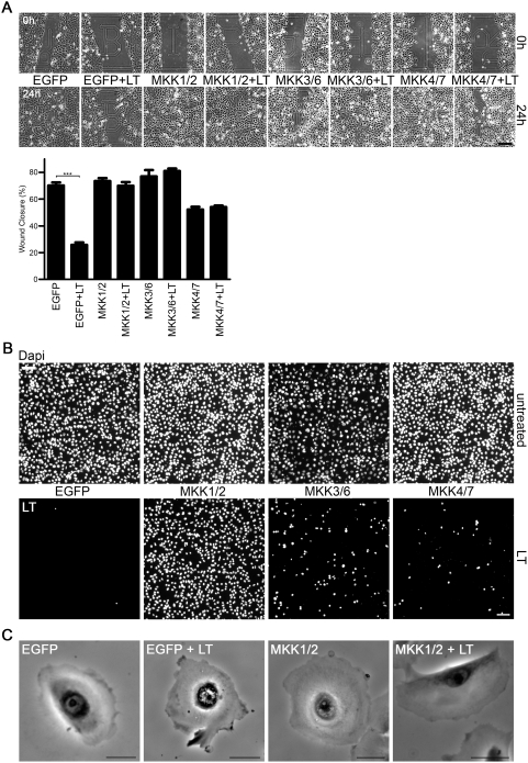
Figure 5. LT-mediated migration defects are MKK-dependent.
(A) Live cell wound healing assay was performed in untreated or LT-treated airway cells (SALE) expressing non-cleavable MKK1/2, MKK3/6 or MKK4/7 mutants or EGFP alone as control. Cells were imaged at time zero and 24 h after the scratch. Scale bar represents 50 µm. Quantification of experiment is shown in lower panel. Data (mean+/−SEM) in each experiment were obtained from 10 different wound areas, n = 3. The overall analysis of variance between all groups was highly significant: F7,72 = 49.6, p<0.0001. (B) Chemotaxis of untreated and LT-treated airway cells expressing non-cleavable MKK1/2, MKK3/6 or MKK4/7 using Boyden chambers (migration for 21 h). Cells on the bottom of the filter were stained with DAPI (blue). Upper panel depicts untreated cells, lower panel LT-treated cells. Scale bar represents 50 µm. (C) A representative frame of bright field live cell movies of untreated and LT-treated airway cells expressing EGFP or non-cleavable MKK1/2-/EGFP after global growth factor stimulation. Scale bar represents 20 µm. Complete movies (S9–12) can be found in Supplemental data.

