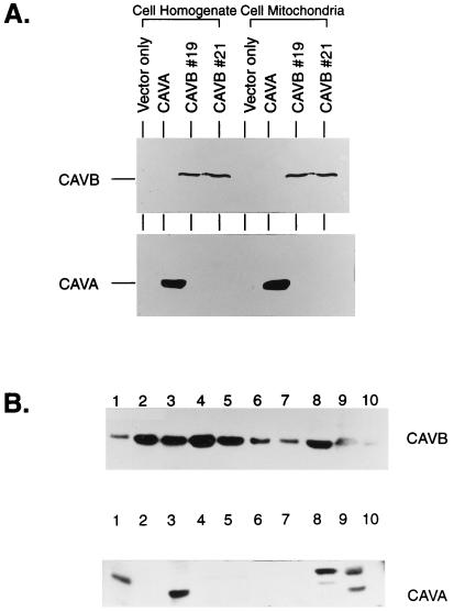
Figure 2.
Expression of CA VB in COS-7 cells and different mouse tissues. (A) The cell homogenate or cell mitochondria from COS-7 cells transfected with vector only, CA VA and CAVB cDNAs clone nos. 19 and 21 were analyzed by SDS/PAGE followed by immunoblotting. The polypeptides for CA VA and CA VB are marked (Fig. 1). (B) Homogenates of different tissues containing 50 μg of protein were analyzed by SDS/PAGE followed by immunoblotting by using anti-mouse CA VB C-tail or CA VA C-tail antisera. Lanes: 1, brain; 2, heart; 3, liver; 4, lung; 5, kidney; 6, spleen; 7, intestine; 8, testis; 9, muscle; 10, pancreas. The polypeptides of 31-kDa mature CA VB were seen in all tissues used here. The polypeptides of 31-kDa mature CA VA and proteolytically nicked 28-kDa polypeptides were seen in few tissues.

