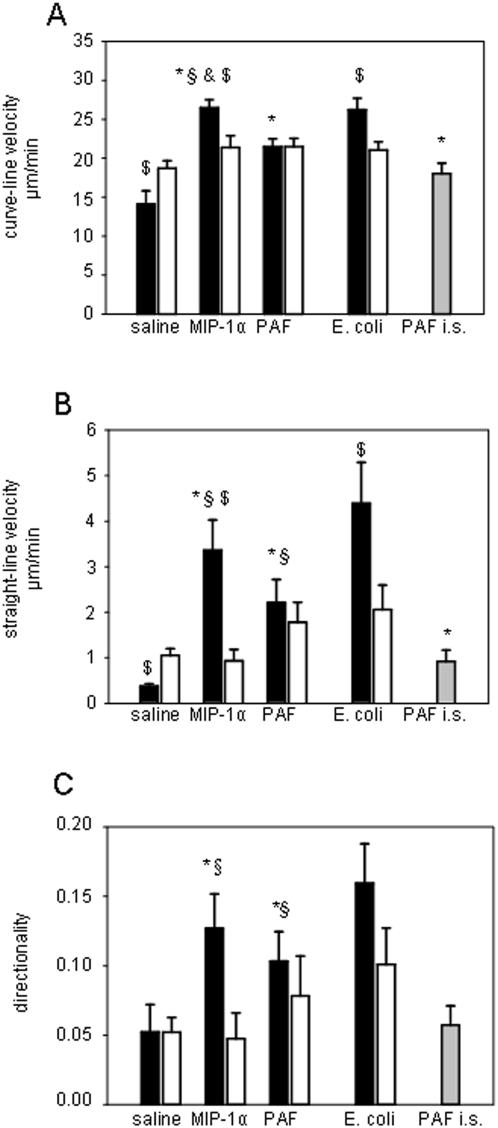
Figure 5. Leukocyte motility.
Parameters of leukocyte motility such as curve-line migration velocity (A), straight-line migration velocity (B), and directionality (C) were determined in digitized in vivo microscopy video sequences 60 min after microinjection of MIP-1α, PAF or E. coli as well as upon intrascrotal application of PAF (PAF i.s.). Interstitially migrating leukocytes (n = 15) were analyzed during 5 min using SimplePCI software. Parameters of leukocyte motility are presented for the vessel side ipsilateral to the microinjection site (black bars), on the contralateral vessel side (white bars), and after intrascrotal application of PAF – (gray bars); mean±SEM; *p<0.05 vs. saline, § p<0.05 vs. PAF i.s., & p<0.05 vs. PAF, $ p<0.05 vs. contralateral side; n = 15; E. coli n = 8.

