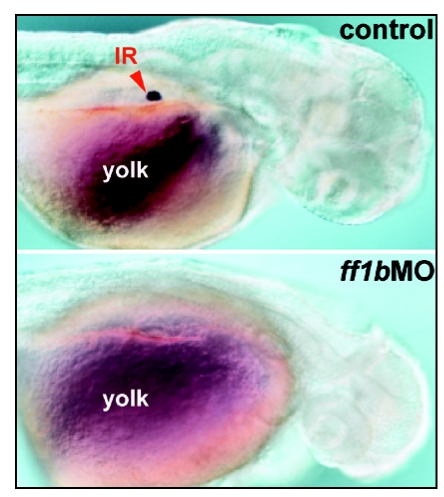
Figure 1.
The detection of steroidogenic interrenal tissue by whole-mount chromogenic 3β-Hsd enzymatic activity assay in wild-type control (upper panel) and ff1b antisense morpholino (ff1bMO) injected (lower panel) embryos. The embryos were treated with 0.003% phenylthiourea from 12 hpf onwards to prevent pigmentation. Dorso-lateral views of embryos at 2 dpf are shown with anterior oriented to the right. The steroidogenic interrenal cells are completely ablated in ff1b morphant. Red arrowhead indicates interrenal tissue (IR), lying above yolk sac in the mid-trunk region.

