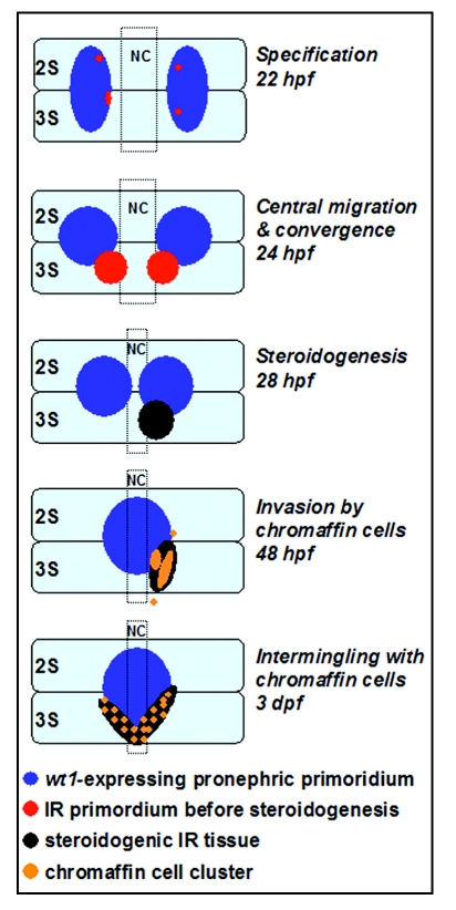
Figure 2.
Early morphogenetic processes in the interrenal development of zebrafish. The parallel migrations of interrenal tissue, pronephros and chromaffin cells in this diagram are depicted based on the results of references 18, 20 and 39. The panels represent dorsal views of embryos, at the indicated stages, oriented with anterior to the top. Through the sequential stages of interrenal specification, migration, steroidogenesis and chromaffin cell invasion, an assembled interrenal organ is evident by 3 dpf. NC, notochord; 2S and 3S, the second and third somite, respectively.

