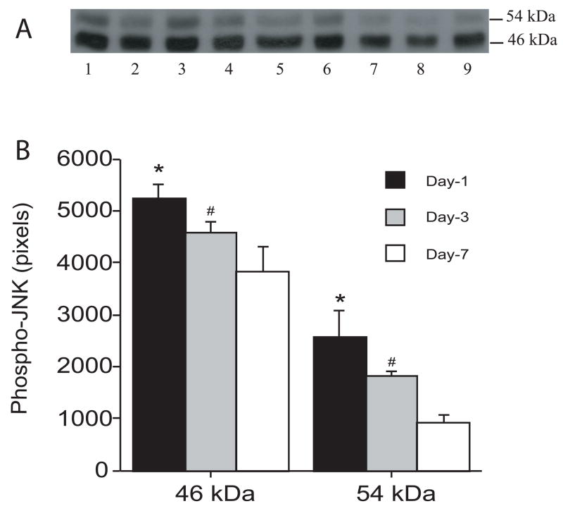
Figure 1.
Time course of the levels of phosphorylated JNK in the ipsilateral basal ganglia after intracerebral infusion of autologous blood. (A) Western blots showing phosphorylated-JNK at 1 day (lanes 1–3), 3 days (lanes 4–6) and 7 days after intracerebral infusion of autologous blood. Equal amounts of protein (50 μg) were used for each sample. (B) Quantification of Western blot analysis. Values are expressed as means ± SD; there are three rats in each group. # p< 0.05 and * p<0.01 vs. day 7.

