
FIG. 6.
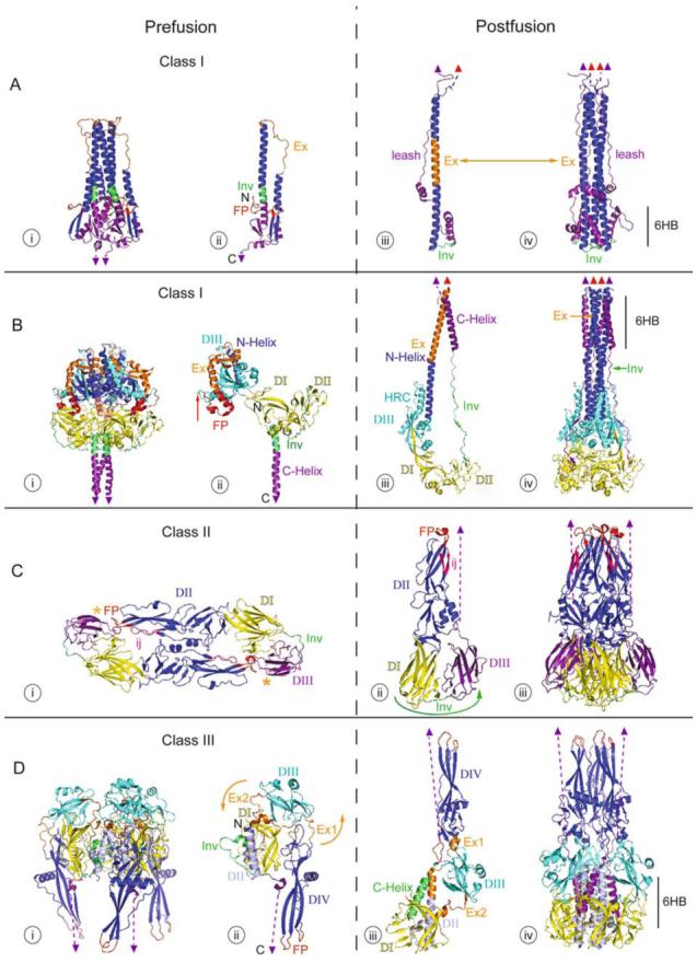
Structures of Class I, II, and III fusion proteins in their pre- and post-fusion forms. The crystal structures of the Class I fusion proteins, Influenza virus HA2 (A) and Paramyxovirus F (B), a Class II fusion protein, TBEV E (C), and a Class III fusion protein, VSV G (D), are shown. The pre-fusion states (i and ii) are on the left and the post-fusion states (iii and iv) are on the right with functional domains identified by color. Fusion peptides are in red, with the domains containing them in dark blue. C-terminal domains, which connect to the virus membrane, are in purple. C-terminal linkers, transmembrane domains, and fusion peptides not visible in the structure are represented by dashed purple lines, purple triangles, and red triangles, respectively. Regions important for the movement of the fusion peptide toward the target membrane are displayed in orange, and regions important for C-terminal inversion, that bring the fusion peptide and transmembrane domains together, are shown in green (in Ai-iii, Bi-iii, Ci-ii, and Di-iii). In the post-fusion trimer structures (iv), the orange and green are replaced by dark blue or purple domain coloring, respectively, to illustrate the similarities among all of the post-fusion forms. Other domains are represented in cyan, yellow, and blue-gray. PDB accession numbers for the structures are: 2HMG (Ai and ii); 1QU1 (Aiii and iv); 2B9B (Bi and ii); 1ZTM (Biii and iv); 1SVB (Ci); 1URZ (Cii and iii); 2J6J (Di and ii); 2CMZ (Diii and iv).