Figure 1.
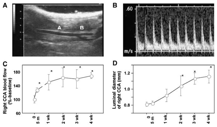
(A) Representative view of the right common carotid artery. Right side is the cephalic side (head), and the left side is the caudal side (tail). A: Right common carotid artery. B: Carotid bifurcation. (B) Representative view of the pulse wave Doppler tracing of the right common carotid artery. (C) Blood flow of the right common carotid artery after flow augmentation. There was an immediate increase in blood flow in the right common carotid artery 5 minutes after the left common carotid artery ligation. Blood flow in the right common carotid artery further increased at 1 week, and the increase was sustained for at least up to 4 weeks. (D) Luminal diameter of right common carotid artery after left common carotid artery ligation. Before surgery to 3 weeks, there was a gradual increase in luminal diameter, which thereafter appeared to plateau. Luminal diameter at 1, 2, 3, and 4 weeks was greater than at baseline (mean±s.d). *P<0.05 compared with baseline (t=0). CCA: common carotid artery.

