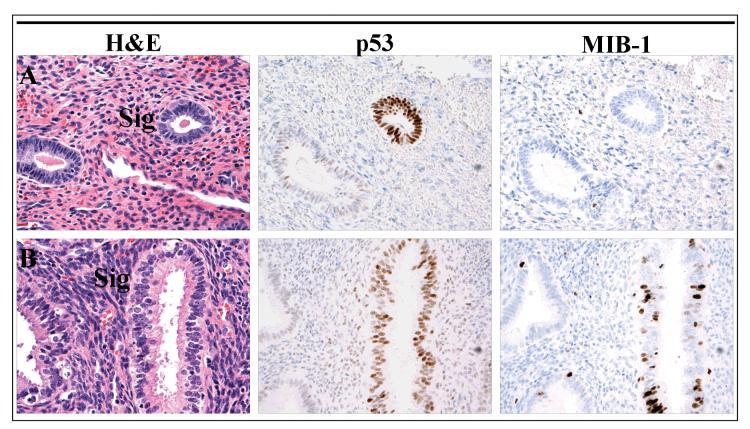
Figure 1.
p53 signatures in benign polyps. Immunostaining for p53 highlights discrete glands which are morphologically indiscernible from the surrounding glands. The proliferative fraction of the p53 signature in Panel A is 0%, similar to an adjacent p53-negative gland, while the signature in Panel B exhibits a higher proliferative index.

