
Fig. 1. Regions of dRN Analyzed for CRF Receptor Immunofluorescence.
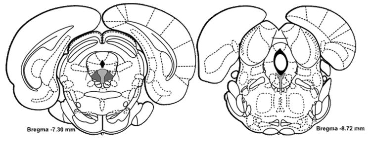
Coronal sections of the dRN illustrating the anterioposterior range of the dRN that were analyzed for CRF receptor immunofluorescence. The dRN was divided into lateral (dark gray fill) and medial (light gray fill) regions for -7.3 mm to -8.0 mm from bregma. At -8.3 mm to -8.72 mm from bregma, only the medial region was analyzed, since the lateral portions of the dRN at this level are not present. Figures adapted from Paxinos and Watson (1997).