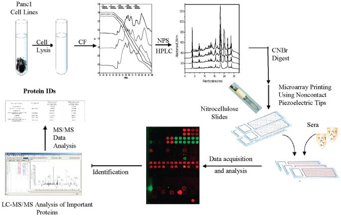
Figure 1.
Overall workflow of the modified protein microarray strategy. Proteins from a cell line/tissue are first extracted and separated in two dimensions (chromatofocusing separated the proteins according to their pI and NPS-RP-HPLC separated them according to their hydrophobicity). Separated fractions are split into three parts. One part is digested with trypsin, one with CNBr, and one is left intact. Intact proteins and CNBr-digested proteins are arrayed on nitrocellulose slides and probed with serum from different stages of disease (in this case, normal, chronic pancreatitis, and pancreatic cancer) to visualize humoral response. Tryptic digests of the spots that showed a differential humoral response were then subjected to protein identification using LC-MS/MS.

