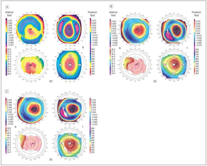
Figure 1.
Orbscan images of study cases. Orbscan image of patient 2 shows central corneal thinning, increased posterior float, and inferocentral corneal steepening (A). Orbscan images of patient 4 at 3 years (B) and 5 years (C) after laser in situ keratomileusis. Both show central corneal thinning, increased posterior float, and central corneal steepening.

