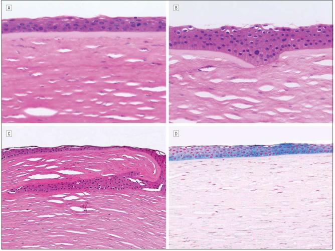
Figure 3.

Cornea with keratectasia from Figure 2 at a higher magnification. A, Central cornea of patient 3 showing intact Bowman layer (hematoxylin-eosin, original magnification ×252). B, Peripheral cornea in patient 3 showing disruption of the Bowman layer at the site of microkeratome incision (hematoxylin-eosin, original magnification ×252). C, Epithelial ingrowth in patient 2 at the point of microkeratome incision (hematoxylin-eosin, original magnification ×126). D, Epithelial iron deposits visualized with Prussian blue stain in patient 2 (original magnification ×126).
