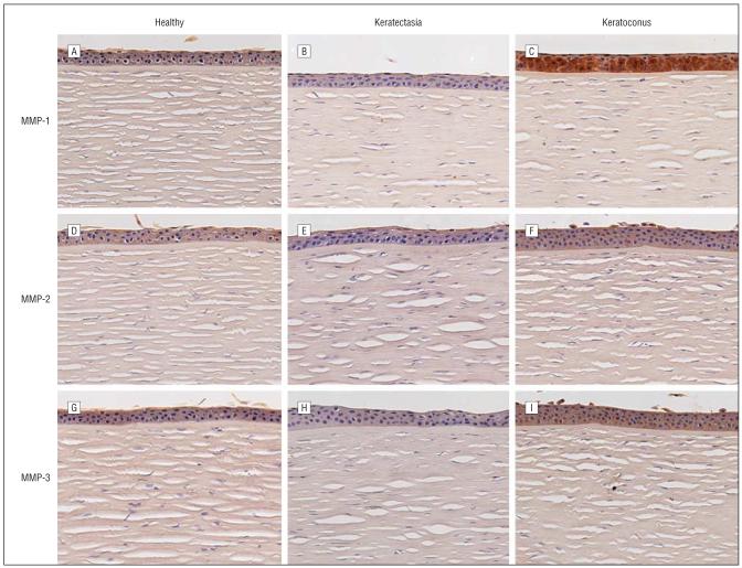
Figure 7.
Immunostaining results for the matrix metalloproteinases (MMPs). A-C, MMP-1 in healthy corneas, corneas with keratectasia, and corneas with keratoconus, respectively. D-F, MMP-2 in healthy corneas, corneas with keratectasia, and corneas with keratoconus, respectively. G-I, MMP-3 in healthy corneas, corneas with keratectasia, and corneas with keratoconus, respectively. Note that the intensity of the MMP-1 staining is very low in the epithelium of the healthy corneas and the corneas with keratectasia. By contrast, strong epithelial staining of MMP-1 is present in the keratoconus case. The MMP-2 and MMP-3 staining was at the background level or absent in healthy corneas, the corneas with keratoconus, and the corneas with keratectasia (chromagen 3,3-diaminobenzidine tetrahydrochloride, original magnification ×126).

