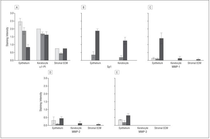
Figure 8.
Semiquantitative immunostaining results for all antibodies. A, Staining intensity for α1-proteinase inhibitor (α1-PI). B, Nuclear staining intensity for Sp1 in corneal epithelial cells and keratocytes in the corneas with keratectasia (open bar, n=5), healthy human corneas (gray bar, n=2), and corneas with keratoconus (black bar, n=2) as scored by 3 masked observers. Staining intensity for matrix metalloproteinase (MMP) 1 (C), MMP-2 (D), MMP-3 (E) in corneal epithelial cells, keratocytes, and stromal extracellular matrix (ECM). Error bars indicate SEM.

