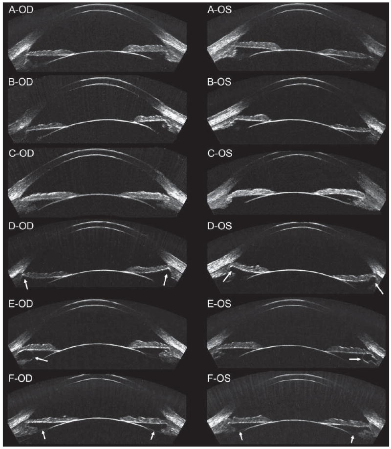
Figure 1.

Geometrically corrected horizontal B-scan images of the anterior segment of right/left pairs in six patients. A) The angle diameter was similar to the sulcus diameter in this patient. B) The angle diameter was smaller than the sulcus diameter in this patient. C) The angle diameter was larger than the sulcus diameter in this patient. D) Anterior segment imaging showed the sulcus to be recessed in this patient (indicated by white arrows). E) Anterior segment imaging found cysts within the ciliary body of each eye (indicated by white arrows). F) The zonules were also visible in this patient (indicated by white arrows).
