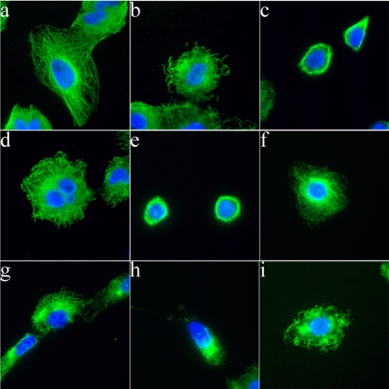
Fig. 3.
Disorazole C1 disrupts microtubules in vivo. Microtubules in A549 cells were partially disrupted after disorazole treatment at 2 nM for 24 h (b) and more completely at 72 h (c) compared with DMSO vehicle control (a). The disruption was similar to that seen with 2 nM vinblastine treatment at 24 h (d) and 72 h (e). A concentration response effect on microtubules after a 1-h treatment was observed with 2 nM (f), 10 nM (g), and 20 nM (h) disorazole C1, similar to 20 nM vinblastine (i). With both compounds, apparent tubulin aggregation was observed at higher concentrations (h and i). Cells were fixed and stained with anti-α-tubulin antibody to visualize microtubules (green) and 4,6-diamidino-2-phenylindole to visualize nuclei (blue).

