
FIGURE 7. Reduced clearance of late-apoptotic cells in trpml mutants Images are by confocal microscopy.
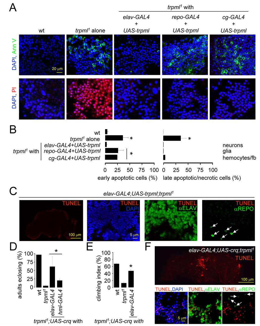
(A) DAPI/Annexin V-FITC and DAPI/PI stained brains from 21 day-old flies.
(B) Percentage of early apoptotic and late apoptotic/necrotic cells in brains from 21 day-old flies. n≥3; *, difference from wt, p≤0.01, ANOVA.
(C) Brains from 21 day-old flies viewed at 310 nm to detect DAPI, 488 nm to detect anti-REPO, 568 nm to detect TUNEL and 633 nm to detect anti-ELAV. Arrows indicate glia.
(D) Percentage of pharate adults without the TM3 balancer. n=3–11; *, difference from trpml1, p≤0.05, ANOVA.
(E) Climbing indices of adult flies. n≥5; *, difference from trpml1, p≤5×10⁻⁵, t-test.
(F) Brains from 21 day-old flies viewed at 310 nm to detect DAPI, 488 nm to detect anti-REPO, 568 nm to detect TUNEL and 633 nm to detect ELAV. Arrows indicate glia.