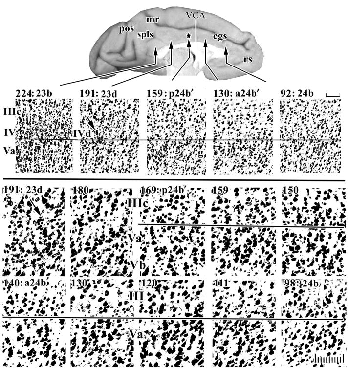
Figure 1.
Systematic sampling of cresyl violet-stained sections through a rhesus monkey case at two magnifications to assess architecture in “b” areas along the cingulate gyrus. Layers in sections for this and subsequent figures are aligned on the border between layers IIIc and Va or layers IV and Va. The star on the surface photograph is the position of the Brodmann border and this “border” is 3 mm caudal to the VCA. The middle cortical layers are magnified to identify changes in small neuron densities in layers IV and Va. The first evidence of a layer IV is the dysgranular layer in area 23d (arrow, #191) and the dysgranular nature of this cortex is emphasized by the fact that layer IIIc/Va pyramids interact at one point, while area 23b caudally has an unbroken layer IV. Higher magnification shows that layer Va neurons in sections #98–130 are relatively large, in #140–159 large pyramids are somewhat smaller, while in #169–180 there are many small neurons in layer Va giving the appearance of a granular layer; this is not layer IV, however. cgs, cingulate sulcus; mr, marginal ramus; pos, parieto-occipital sulcus; rs, rostral sulcus; spls, splenial sulci; scale bars, 100 μm.

