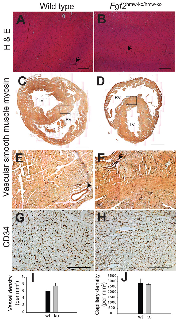
Figure 3. Fgf2hmw-ko/hmw-ko mice (2 month old) have normal myocardial architecture, blood vessel and cardiac capillary density.
(A,B), H & E stained cross-sections of wild type (A) and Fgf2hmw-ko/hmw-ko (B) mouse hearts. (C,D), SMC myosin immunohistochemistry on wild type (C) and Fgf2hmw-ko/hmw-ko (D) hearts indicating SMC containing blood vessels. Images of the whole heart are reconstructed by combining several smaller images (10×1.6 objective magnification) using Adobe Photoshop. Area under the box in (C) and (D) is magnified and represented by (E) and (F), respectively. (G,H), CD34 immunohistochemistry on wild type (G) and Fgf2hmw-ko/hmw-ko (H) hearts depicting cardiac endothelial capillaries. Note that myocardium and myocardial capillarogenesis (arrows, B) is normal in Fgf2hmw-ko/hmw-ko mice. Also, the distribution of SMC containing blood vessels (arrows, D,E) and their quantification (I) is not significantly different between the wild type and Fgf2hmw-ko/hmw-ko mice (P = 0.1705). In addition, there is no qualitative (H) and quantitative (J) difference in CD34-stained capillaries between the wild type and Fgf2hmw-ko/hmw-ko mice (P = 0.7877). Scale bar: 200 µm (A,B), 1 mm (C,D), 100 µm (G,H). RV, right ventricle; LV, left ventricle.

