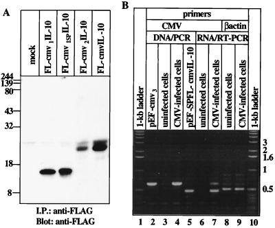
Figure 2.
cmvIL-10 expression. (A) Western blotting analysis of COS-1 cell-conditioned media. COS-1 cells were transiently transfected with the pEF-SPFL [lane 1 (mock)], the pEF-SPFL-cmv1 [lane 2 (FL-cmv1IL-10)], the pEF-SPFL-cmv1SP [lane 3 (FL-cmv1SPIL-10)], the pEF-SPFL-cmv2 [lane 4 (FL-cmv2IL-10)], or the pEF-SPFL-cmvIL-10 [lane 5 (FL-cmvIL-10)] expression vectors. Three days later, 1 ml of the conditioned media was subjected to immunoprecipitation and Western blotting experiments with anti-FLAG antibody. The molecular weight markers are shown on the left. (B) CMV-infected cells express cmvIL-10. PCR (lanes 3 and 4) or RT-PCR (lanes 6 and 7) with the same sets of primers was performed with DNA or RNA isolated from virus-infected (lanes 4 and 7) or uninfected (lanes 3 and 6) cells as described in Materials and Methods. Plasmids pEF-cmv3 (lane 2) and pEF-SPFL-cmvIL-10 (lane 5) were used for PCR as positive controls. A 1-kb ladder was run in lanes 1 and 10.

