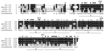
Figure 3.
Alignment of amino acid sequences of human IL-10 and its viral homologs. The alignment of the amino acid sequences of cellular IL-10 encoded by the human genome (23) and viral IL-10s encoded by EBV [ebvIL-10 (10)], OV [ovIL-10 (12)], and CMV [cmvIL-10 (this study)] are shown. A consensus sequence is shown on the bottom. Identical amino acids corresponding to the consensus sequence are shown in black outline with white lettering. Similar amino acids are shown in gray outline with white lettering. Amino acid residues are numbered starting from first Met residue (signal peptide amino acids are included). The α-helices A through F, taken from the crystal structure of IL-10 and ebvIL-10 (30, 36), are underlined. Symbols: ① and ② designate Cys residues of IL-10 that form two intramolecular disulfide bridges (30). Asterisks denote amino acids predicted to be involved in interaction with IL-10R1 (32). The bold asterisks represent those residues involved in the interaction with IL-10R1 that are conserved among all the IL-10s. ■ points to conserved amino acids within regions involved in interaction with IL-10R1. □ points to conserved amino acids in the middle of IL-10 homologs that may be involved in interaction with IL-10R2. Arrows indicate positions of introns within IL-10 and cmvIL-10 genes. Numbers in parentheses represent the number of introns in IL-10 and cmvIL-10 (intron number within IL-10/intron number within cmvIL-10); the minuses at (2/−) and (4/−) denote no intron in cmvIL-10 at these positions. The triangle represents the position of Ala-98 of ebvIL-10. The program pileup of the Wisconsin Package, Version 9.1, Genetics Computer Group, Madison, WI, was used with the following parameters: the gap creation penalty 10, the gap extension penalty 2. The boxshade 3.21 program was used for shading of the alignment file.

