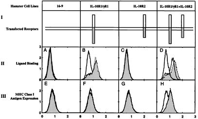
Figure 4.
Ligand binding and MHC class I antigen induction. (row I) Schematic of four cell lines used in these experiments: the parental Chinese hamster 16–9 cells and three 16–9-based cell lines expressing human IL-10R1/γR1 chimeric receptor or human IL-10R2 alone or both receptors together, which were created and described in detail (21). (Row II, A–D) The cells described in row I were incubated for 30 min at 4°C with conditioned medium from COS-1 cells transfected with one of the following plasmids: the control vector pEF-SPFL (open areas, thick lines); the pEF-SPFL-IL-10 (open areas, thin lines), or the pEF-SPFL-cmvIL-10 (shaded areas, thin lines). Ligand binding to the cell surface was determined by flow cytometry with anti-FLAG antibody (Sigma) as the primary antibody and FITC-conjugated goat anti-mouse IgG (Santa Cruz) as the secondary antibody. Here and in row III, the ordinate represents relative cell number, and the abscissa is relative fluorescence. (Row III, E–H). The ability of IL-10 and cmvIL-10 to induce MHC class I antigen expression was demonstrated by flow cytometry as described (21). The cells described in row I were left untreated (open areas, thick lines) or treated with conditioned media (100 μl) from COS-1 cells transfected with the pEF-SPFL-cmvIL-10 plasmid (shaded areas, thin lines) or with Hu-IL-10 (100 units/ml; open areas, thin lines).

