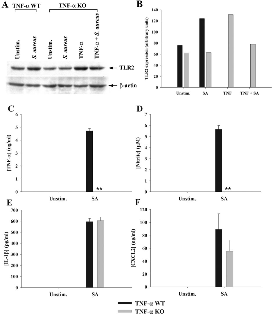
Figure 3. Astrocytes produce TNF-α in response to S. aureus that functions in an autocrine/paracrine manner to increase TLR2 and NO expression.
Primary astrocytes isolated from TNF-α WT or KO mice were stimulated with heat-inactivated S. aureus +/− 100 ng/ml of recombinant mouse TNF-α for 24 h, whereupon whole cell extracts were prepared and analyzed for TLR2 expression by Western blotting (A and B). Results are presented as the raw gel data (A) and quantitative analysis of TLR2 expression by densitometry (B). For quantitation in (B), the pixel intensity of each TLR2 band was normalized to the amount of actin to verify uniformity in gel loading. In addition, cell-free supernatants were collected at 24 h following bacterial exposure and analyzed for TNF-α (C), nitrite (D), IL-1β (E), and CXCL2 (F) production. Results are reported as the mean ± SD of three independent wells for each experimental treatment (C–F). Significant differences between TNF-α WT and KO astrocytes stimulated with S. aureus are denoted with asterisks (**, p < 0.001). Results are representative of two (A–B) or six (C–F) independent experiments.

