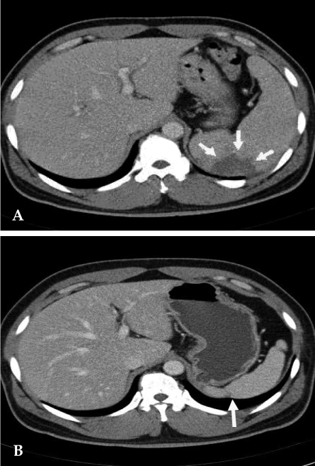
Fig. 1.
(A) Contrast-enhanced abdomen CT scan reveals partial splenic abscess that is a wedge shape (arrow) and splenomegaly. (B) Twelve months later, follow-up CT scan reveals normalized spleen size of about 11 cm compared with the previous splenomegaly size of 15 cm. A portion of the splenic abscess changed into cortical dimpling (arrow) with segmental shrinkage.

