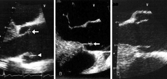
Fig. 2.
(A) Transesophageal echocardiography reveals one vegetation (4 × 2 mm, arrow) on the right aortic coronary cusp and the other vegetation (5 × 3 mm, arrow head) on the noncoronary aortic cusp upon admission. (B) Two months later, follow up transesophageal echocardiography (TEE) reveals no interval change of the vegetation (arrow) size within the noncoronary cusp, but vegetation within the right coronary cusp was completely resolved. (C) Seven months later, follow up TEE shows complete resolution of the remained vegetation.

