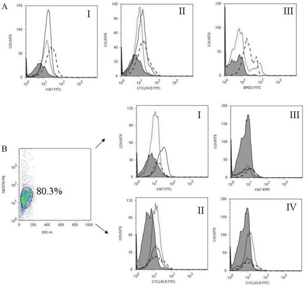
Figure 3.

CD133+ retinal cells expression of proliferation markers following culture with FGF/N2 and FGF-LIF overnight. CD133+ retinal cells following culture overnight with either FGF/N2 or FGF-LIF were analysed by flow cytometry for the expression of the proliferation markers Ki67, Cyclin D1 or BrdU incorporation.(A) Histograms show the expression of Ki67 (AI) and Cyclin D1 (AII) after culture with FGF/N2 (dotted line) or FGF-LIF (dashed line) compared to expression immediately following CD133+ retinal cell isolation (black line) against isotype control (Grey filled peaks). LIF within cultures causes an increase in expression of cell proliferation markers. Figure AIII shows incorporation of BrdU within cells following culture overnight with FGF/N2 (dotted line) or FGF-LIF (dashed line) indicating the generation of newly formed cells. (B) Dot plot demonstrates gated CD133+ Nestin+ events presented as mAb fluorescence against side scatter. CD133+Nestin+ and Nestin-CD133+ retinal cells were analysed for coexpression of Ki67 (I, III) or Cyclin D1 (II, IV). At time 0, Nestin positive cells expressed higher levels of proliferation markers Ki67 (I 5.90%, MFI 19.91) and Cyclin D 1 (II 12.01%, MFI.22.11) than nestin negative cells (Ki67 III Ki67 2.10%, MFI 11.19 and Cyclin D 1 IV 9.50%, MFI 19.07). Purified cells directly ex vivo (black line), culture overnight with FGF/N2 (dotted line) or FGF-LIF (dashed line).
