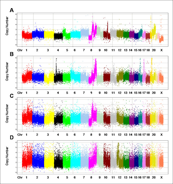
Figure 2.
Copy number data for 4 samples of varying 2p-RSEs. In each panel, markers are arranged along the chromosomes and colored by chromosome. Chromosomes are typically labeled; some of the smaller chromosomes are unlabeled due to space constraints. The X axis represents chromosomes in genomic order of markers; the Y axis is the absolute copy number measurement. (A) shows one of the best samples with 2p-RSE = 0.109. (B) shows an average "high quality" sample with 2p-RSE = 0.147. (C) shows a sample that just fails to meet the high quality threshold with 2p-RSE = 0.184. (D) shows the worst passed sample with 2p-RSE = 0.247.

