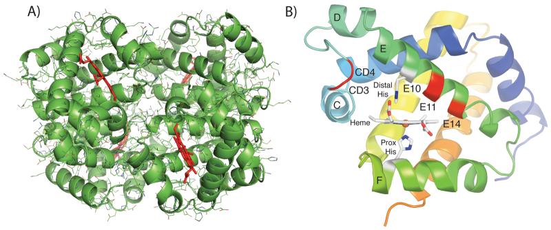
Figure 2.
(A) Tetrameric structure of bovine Hb at pH 5.7 (heme groups are shown in red). (B) Highlighted amino acid differences in the E helix and CD turn. The structure of the trout IV β subunit is shown. The amino acid residues listed in Table VI are highlighted in red and labeled. The C, D, E, and F helices and the CD turn are labeled along with the heme and proximal and distal histidine residues.

