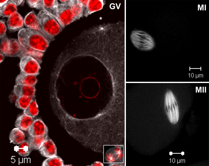
Fig. 1. Activated SFKs distribute in microtubule-like patterns at the GV, MI and MII stages of meiosis.
Germinal vesicle stage oocytes were collected from PMSG primed mouse ovaries and matured in vitro for 0, 8 or 16 hours followed by fixation. Oocytes were labeled with a monoclonal antibody against activated Src-family PTKs (clone 28) and detected with Alexa-488-goat anti-mouse IgG (white) and co-labeled with the DNA dye Hoechst 33258 (red). Active SFKs are distributed in microtubule-like patterns consistent within oocytes and companion cumulus cells; see mitotic spindle microtubule labeling in cumulus cell (GV inset). Within GV stage oocytes, SFKs localized to cytoplasmic microtubule arrays surrounding the nuclear envelope as well as cortical microtubule organizing centers (*). At MI and MII, SFKs localize primarily to microtubules of the metaphase spindle.

