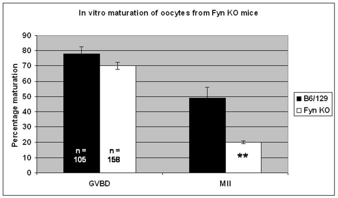
Fig. 7. Oocytes from FYN (−/−) mice exhibit reduced meiotic potential.
Oocytes and COC from FYN (−/−) and wildtype controls collected at the GV stage were matured in vitro for 16hrs and subjected to fluorescence analysis as before. FYN (−/−) oocytes exhibited reduced meiotic potential as indicated by the fact that while FYN (−/−) oocytes underwent GVBD to comparable levels as wildtype controls (70 % versus 76%, respectively), significantly fewer FYN (−/−) oocytes achieved MII (20% versus 49%; **P<0.05).

