
Fig. 1.
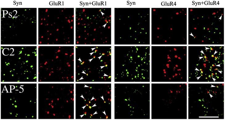
Confocal images of abducens motor neurons showing punctate staining for the presynaptic marker synaptophysin (Syn; green), GluR1 AMPAR subunits (red), or GluR4 AMPAR subunits (red) from each of the conditioned groups examined: pseudoconditioned for two sessions (Ps2), conditioned for two sessions (C2), and conditioned during application of AP-5 for two sessions (AP-5). Colocalization of AMPAR with synaptophysin punctate staining is also shown (GluR1+Syn, GluR4+Syn) and indicated by the arrowheads. Scale bar = 2 μm.