
Fig. 2.
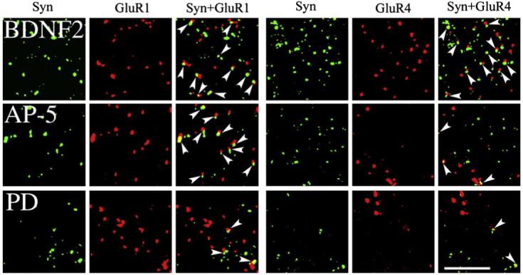
Confocal images of punctate staining of abducens motor neurons for synaptophysin, GluR1, and GluR4 AMPAR subunits from each of the BDNF-treated groups examined: BDNF application for two sessions (BDNF2), coapplication of BDNF with AP-5 for two sessions (AP-5), and coapplication of BDNF with PD98059 for two sessions (PD). Colocalization of AMPAR with synaptophysin punctate staining is also shown (GluR1+Syn, GluR4+Syn) and indicated by the arrowheads. Scale bar =2 μm.