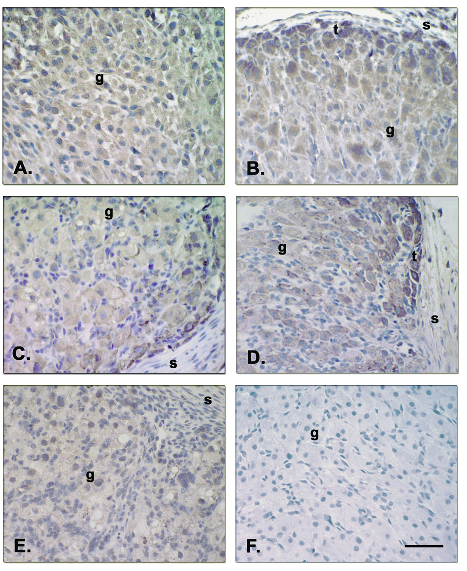
Figure 6.
Immunostaining for FASL and FAS in CL at various stages of the luteal lifespan during the natural menstrual cycle [FASL at ECL (Panel A), MCL (Panel B), LCL (Panel D) and VLCL(Panel E), and FAS at LCL (Panel C)]. Both FASL and its receptor followed a similar pattern of specific staining where theca (t) luteal cells appeared more intensely stained than the granulosa (g) cells by the MCL to LCL stages. Staining was not found in the negative control (FASL Ab preabsorption, Panel F) nor in the endothelial cells or stroma cells (s). Scale bar 50 µm.

