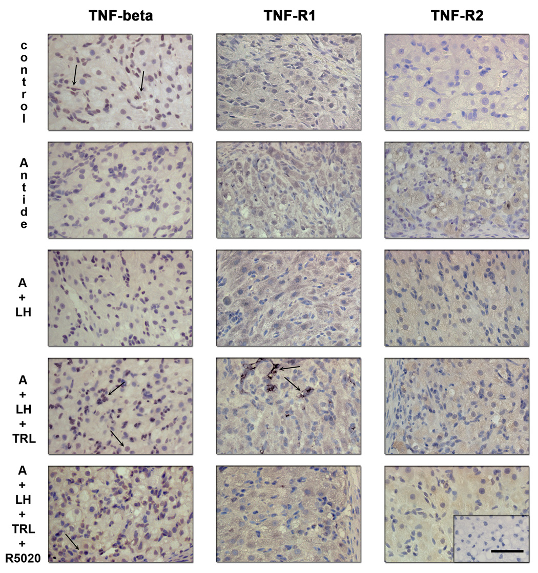
Figure 8.
Immunohistochemistry for TNF-β, TNF-R1 and TNF-R2 following gonadotropin and/or steroid ablation and replacement. Controls (untreated, MLCL) exhibited staining comparable to that described in detail in previous Figure 4 and Figure 5. Specific staining for TNF-β (left panels) was detected in the cytoplasm and, in some cases, in the cell nucleus of the granulosa, theca luteal cells and endothelial cells (arrows). Immunostaining in the luteal endothelial cells appeared to decrease after antide (A) treatment and LH failed to restore its expression. Trilostane and R5020 treatments appeared to restore its expression. Immunostaining for TNF-R1 (center panels) was found in the granulosa and theca luteal cells, but not in endothelial cells and displayed little change in expression, except for an increase in discrete groups of luteal cells after trilostane treatment. TNF-R2 protein expression (right panels) was also found in the granulosa and theca luteal cells and appeared to increase after antide treatment. LH prevented this increase and Trilostane treatment was able to mimic in part the antide effect. Scale bar 50 µm.

