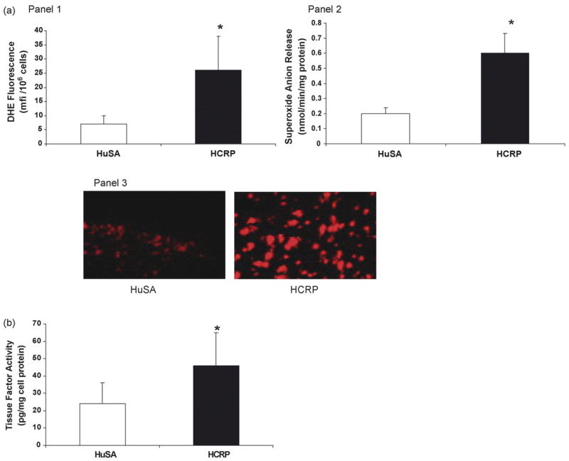
Fig. 1.
(a) Effect of CRP on superoxide anion release in rat macrophages: HCRP/HuSA (20 mg/kg bw for 3 days, n = 5 in each group) was injected in the peritoneal cavity of the rats. The rats were sacrificed on the 4th day and peritoneal macrophages were isolated. Superoxide anion release was assessed by DHE fluorescence (Panel 1) and ferricytochrome C reduction (Panel 2) as depicted in Section 2. Panel 3 is a representative picture of macrophages loaded with DHE. The results are mean ± S.D. of five different experiments and are expressed as per mg cell protein. *p < 0.01 compared to HuSA. (b) Effect of CRP on tissue factor activity in rat macrophages: HCRP/HuSA (20 mg/kg bw for 3 days, n = 5 in each group) was injected in the peritoneal cavity of the rats. The rats were sacrificed on the 4th day and peritoneal macrophages were isolated. Tissue factor activity was assessed as described in Section 2. *p < 0.05 compared to HuSA.

