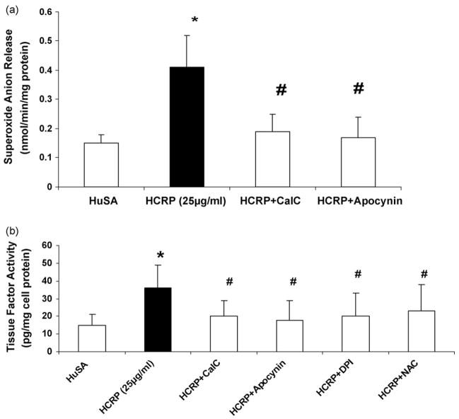
Fig. 2.
(a) Effect of inhibitors to PKC (10 μM calphostin C), NADPH oxidase (100 μg/mL apocynin), on superoxide anion release and (b) effect of inhibitors to PKC (10 μM calphostin C), ROS scavengers (NAC, 10 μg/mL; DPI, 10 μM; apocynin, 100 μg/mL), on tissue factor activity in pouch macrophages. Inhibitors were injected into sterile air pouches of Wistar rats (n = 3 in each group) at least 2 h prior to administration of HCRP (25 μg/mL) or HuSA and superoxide and tissue factor were assessed as described in Section 2. The results are mean ± S.D. *p < 0.05 compared to HuSA and #p < 0.05 compared to HCRP.

