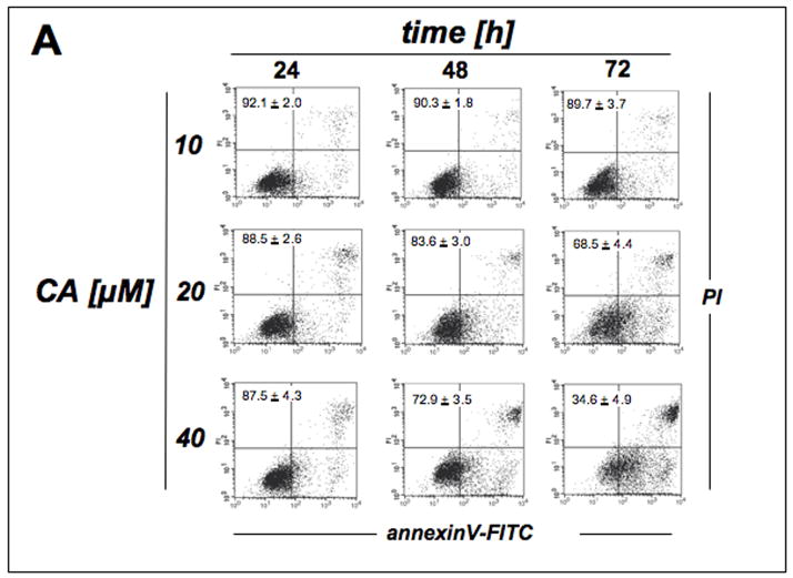
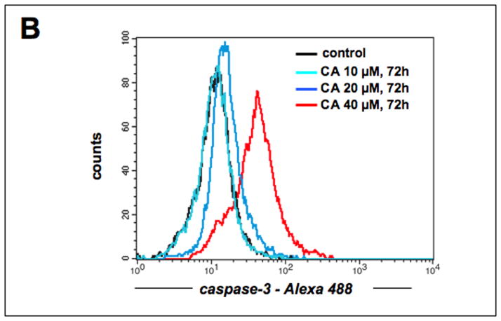
Figure 4. Prolonged exposure to cinnamic aldehyde dose-dependently induces apoptosis with procaspase-3 cleavage in human A375 melanoma cells.
(A) Time course (24, 48, and 72 h) of induction of cell death by exposure to increasing doses of CA (10, 20, and 40 μM) was assessed by flow cytometric analysis of annexinV-FITC/propidium iodide-stained cells. The numbers indicate viable cells (AV−, PI−, lower left quadrant) in percent of total gated cells (mean ± SD, n=3). (Viability of untreated controls was 92.1 ± 1.0 %; data not shown). (B) CA-induced (10, 20, and 40 μM, 72 h) caspase-3 activation was examined by flow cytometric detection using an Alexa Fluor 488-conjugated monoclonal antibody against cleaved procaspase-3. One representative experiment of three similar repeats is shown.


