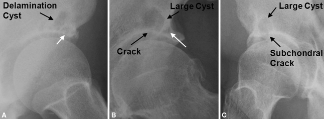
Fig. 1A–C.
(A) A frog lateral radiograph of the hip shows an example of an adjacent type of delamination cyst. The delamination cyst is located directly adjacent and medial to a lateral acetabular cyst (white arrow). (B) An AP radiograph of the hip shows an example of an adjacent type of delamination cyst (white arrow denotes the adjacent lateral acetabular cyst) with an associated crack in the acetabular subchondral bone. The crack extends from the hip into the cyst. (C) This frog lateral radiograph of the hip shows an example of a cracked type of delamination cyst. The delamination cyst is a large, isolated cyst with an associated crack in the acetabular subchondral bone extending from the hip into the cyst.

