Figure 3.
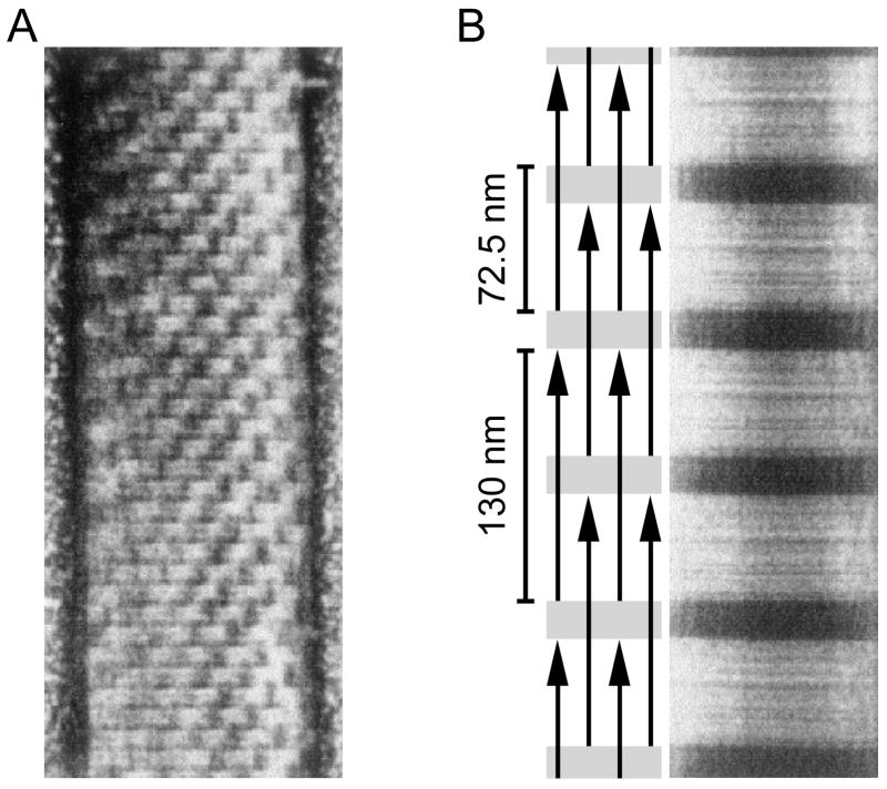
Explanation of large scale (72 nm) structures often present in large diameter mollusc thick filaments. A. A mollusc thick filament with a checkerboard pattern. B. A reconstituted mollusc paramyosin filament with a simple light-dark banding pattern in which the distance of one repeat unit (one dark and one light band) is 72.5 nm. Schematic shows how an overlap-gap binding of individual paramyosin molecules explains the observed staining pattern (only the gap portions take up the stain). Modified from Cohen (1998).

