Figure 5.
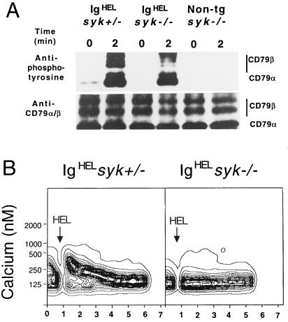
Syk is not required for antigen-induced phosphorylation of CD79α and CD79β but is essential for a calcium response. (A) Western blot of immunoprecitated CD79β from lysates of syk−/− and syk+/− IgHEL and syk−/− nontransgenic bone marrow cells. Cells were stimulated with HEL (1 μg/ml) for 2 min or left unstimulated. Panels show induced tyrosine phosphorylation by probing the immunoprecipitates for antiphosphotyrosine (4G10), relative to the total immunoprecipitated protein measured by reprobing the same blot with antisera to CD79α and β. Results are representative of three experiments. (B) Flow cytometry of indo-1-loaded cells was used to monitor intracellular calcium levels as a function of time after stimulation of IgHEL syk−/− and syk+/− B cells from bone marrow. All measurements were gated on immature, B220lo cells. Arrows indicate time of addition of HEL antigen (1 μg/ml). Equivalent results were obtained from three separate experiments.

