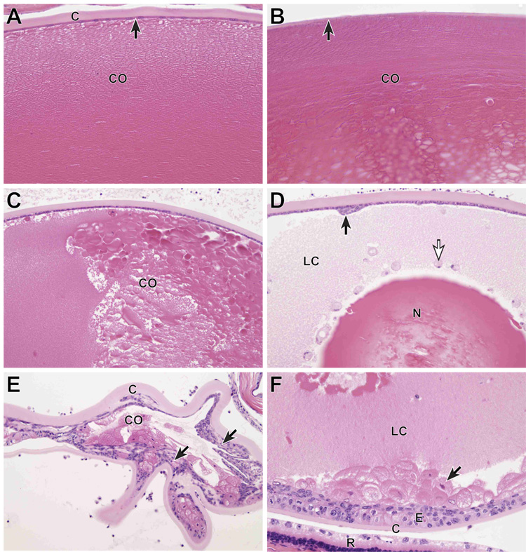
Figure 4.
Ocular histology. A) Normal adult FVB/NJ +/+ mouse. The anterior lens capsule (C) is acellular and eosinophilic. Directly beneath the capsule is a monolayer of cuboidal lens epithelium (arrow). The lens fibers in the anterior lens cortex (CO) are tightly packed and clearly delineated. The lens fibers are the remnants of lens cells and make up the bulk of the adult lens. B) Normal adult FVB/NJ +/+ mouse. The posterior lens capsule (arrow) is normally much thinner than the anterior lens capsule and lens epithelial cells are absent. The posterior lens cortex has a subtle lamellar structure and is more dense that the anterior cortex. C) K14HPV18E7 Tg mouse, 6 months old. The anterior lens capsule and underlying lens epithelium are normal, but the orderly structure seen in the WT mouse is absent and the cortex (CO) has developed a globular appearance, representing masses of degenerating lens fibers. D) K14HPV18E7 Tg mouse, 6 months old. The anterior lens capsule (C) is normal, but a focal patch of reactive proliferation of the lens epithelium is present (arrow). Much of the lens cortex has become liquefied (LC) and some of this degenerating cortical material is contained inside free-floating lens epithelial cells (white arrow). The lens nucleus (N) remains, but centrally shows signs of fragmentation. E) K14HPV18E7 Tg mouse, 8 months old. In this severely degenerated lens, the anterior capsule (C) is thickened and throughout the lens there is massive anterior and posterior proliferation of the lens epithelium (arrows). Small amounts of degenerating cortex (CO) remain. F) K14HPV18E7 Tg mouse, 6 months old. Although lens epithelium is normally absent beneath the posterior lens capsule (C), here there is extensive lens epithelial proliferation (E) of lens epithelium of normal appearance as well as a large number of “bladder” cells. The posterior lens cortex is liquefied (LC). A portion of the degenerated retina (R) with changes typical of Pde6brd1 is present.

