Fig. 3.
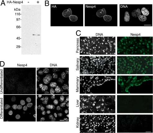
Nesp4 is expressed in secretory epithelial cells. (A) Western blot analysis of HeLa cells using rabbit anti-Nesp4. The cells were either nontransfected (−) or expressed HA-Nesp4 (+). A single band (≈54 kDa) corresponding to HA–Nesp4 is evident only in the transfected sample. The relatively slow migration of HA–Nesp4 (its predicted molecular mass is 42 kDa) is likely caused by its high proline content (9%). (B) HeLa cells expressing HA–Nesp4 and labeled with rabbit anti-Nesp4 and mouse anti-HA. The rabbit anti-Nesp4 only decorates the NE of transfected cells (C) Immunofluorescence microscopy of mouse tissue cryosections using the rabbit anti-Nesp4 antibody. (D) Immunofluorescence microscopy of both undifferentiated and differentiated HC11 mouse mammary cells using the anti-Nesp4 antibody. In all positive cells Nesp4 is localized at the NE. All specimens were stained with Hoechst dye to reveal the DNA. (Bars: B and D, 10 μm; C, 40 μm.)

