Fig. 3.
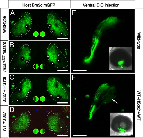
Cell–cell signaling through Radar is sufficient for ventral tectum innervation. (A–D) Rescue experiment. Brn3c:mGFP-labeled retinotectal projections were imaged in vivo at 7 dpf. The tectal neuropil is outlined with a dashed line. Green-filled circles summarize dorsal and ventral tectum innervation results. (A) WT tecta show full innervation. (B) radars327 mutants lack ventral innervation. (C) radarWT expression from heatshock-promoter rescues the retinotectal phenotype of the mutant. In the case shown, only one side was rescued. PCR-based genotyping confirmed that only the rescued eye contained hsp70:radarWT; the other eye had likely not received the injected plasmid because of the mosaicism inherent in transient transgenesis. (D) The radar gene acts cell-nonautonomously in the retina. WT cells transplanted into radars327 host embryos are sufficient to rescue ventral innervation. Only the host carried the Brn3c:mGFP transgene. Donor-derived cells (blue arrowheads) were labeled with rhodamine dextran, and do not contribute to the tectum. (E and F) Gain-of-function experiment. DiO was injected into the ventral retina, and its labeling pattern was imaged from a lateral view. Insets show injected eye. In normal WT larvae (E), ventral RGCs project exclusively to the dorsal tectum. In chimeric WT larvae (F) that have received a transplant of WT cells carrying the hsp70:radarWT construct, some ventral axons ectopically innervate the ventral tectum (arrow). Asterisks (in A–D) show positions of skin melanophores. (Scale bars, 100 μm.)

